INTRODUCTION
Hi one and all. Welcome to our blog!!! Don’t you guys think that it is so pretty and nice? I bet you all do. Anyway, we believed you guys will have an interesting and enriching time when visiting our blog. By visiting our blog, you all will be able to know more about one of our sense organs --- the eye. Over here, we will give you a brief introduction about our wonderful eye and also at the same time let you guys know some interesting facts and viewpoints.
Our amazing sense organ—THE EYE

Have you guys ever wondered how does our eye work? Why can we see things? Well, if you continue reading, you will be able to find out why. Eyes are organs that detect light. Different kinds of light-sensitive organs are found in a variety of animals. In the human eye, light enters the pupil and is focused on the retina by the lens. Light-sensitive nerve cells called rods (for brightness) and cones (for color) react to the light. They interact with each other and send messages to the brain that indicate brightness, color, and contour.
When you look at an object, light rays are reflected from the object to the cornea, which is where the miracle begins. The cornea is a transparent structure found in the very front of the eye that helps to focus incoming light.
Behind the cornea is a colored ring-shaped membrane called the iris. The iris has an adjustable circular opening called the pupil, which can expand or contract depending on the amount of light entering the eye. A clear fluid called the aqueous humor fills the space between the cornea and the iris.

Situated behind the pupil is a colorless, transparent structure called the lens. The lens' job is to make sure the rays come to a sharp focus on the retina. The resulting image on the retina is upside-down. Here at the retina, the light rays are converted to electrical impulses which are then transmitted through the optic nerve, to the brain, where the image is translated and perceived in an upright position!
Ciliary muscles surround the lens. The muscles hold the lens in place but they also play an important role in vision. When the muscles relax, they pull on and flatten the lens, allowing the eye to see objects that are far away. To see closer objects clearly, the ciliary muscle must contract in order to thicken the lens. This is called accommodation.
The interior chamber of the eyeball is filled with a jelly-like tissue called the vitreous humor. After passing through the lens, light must travel through this humor before striking the sensitive layer of cells called the retina. The retina is the innermost of three tissue layers that make up the eye. The outermost layer, called the sclera, is what gives most of the eyeball its white color. The cornea is also a part of outer layer.
The middle layer between the retina and sclera is called the choroid. The choroid contains blood vessels that supply the retina with nutrients and oxygen and removes its waste products. It is pigmented black so as to prevent internal reflection of light.

Embedded in the retina are millions of light sensitive cells, which come in two main varieties: rods and cones. Rods are good for monochrome vision in poor light, while cones are used for color and for the detection of fine detail. Cones are packed into a part of the retina directly behind the retina called the fovea. When light strikes either the rods or the cones of the retina, it's converted into an electric signal that is relayed to the brain via the optic nerve. The brain then translates the electrical signals into the images we see.
Wow, don’t you think that’s amazing? Read up on our various blog posts to know more about the eye! (:
Extraocular Implants
Monday, March 3, 2008/
11:41 PM
The ophthalmologist is back once again! :D
If corneal tattoos are not enough for you, you might be shocked to know that you can 'hang' little decoratives on the whites of your eyes, just like how you put them on your ears.
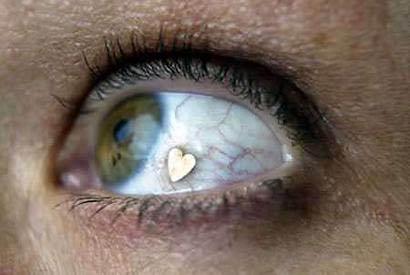
Extraocular implants, also simply known as eyeball jewelry, is not only possible, but legal in Netherlands, as long as its performed by a legalised ophthalmologist (Ahem, that's me) under sterile conditions.
In 2002 the Netherlands Institute for Innovative Ocular Surgery developed a cosmetic extraocular implant (CEI) that can be implanted within the superficial, interpalpebral conjunctiva. The implant does not interfere with the ocular functions, ie the visual performance and motility. The CEI is made of a specially designed material that can be molded in all kinds of desired shapes and sizes.
This is a short description of a CEI implantation under eye-drop anesthesia: - The eye is anesthesized with routine eye-drop anesthesia medication.
- The eye is decontaminated and draped with routine techniques used in ocular surgery.
- An INTRAconjunctival bleb is created by injection of BSS in the most superficial conjunctival layer. The bleb may be positioned adjacent to the temporal limbus.
- With conjunctival scissors, the bleb is opened and a superficial intraconjunctival tunnel is made.
- The tunnel is checked to determine if it is wide enough to accomodate the CEI implantation device.
- The CEI is then loaded into the CEI implantation device, and the device is inserted into the tunnel up to the desired final position of the CEI.
- Using a routine fine-forceps, the CEI is removed from the CEI implantation device and the CEI implantation device is withdrawn from the conjunctival tunnel.
- With a spounge, all excess BSS is removed from the implantation area.
- At the end of the procedure, antibiotic drops are applied onto the eye.
- Postoperative management includes antibiotic eyedrops for 1-2 week(s).
According to the NIIOS, patient satisfaction is high and no side effects of the treatment have been noticed with a follow-up of more than one year.
----
Here is the end of my post and I hope you enjoyed learning about eye modifications. Some of you may feel disgusted, some may feel that it's cool, but I strongly urge everyone of you to think twice before undergoing any of these treatments. Think before you act!! :)
(Done by: Wong May Ping 24/4F)
